
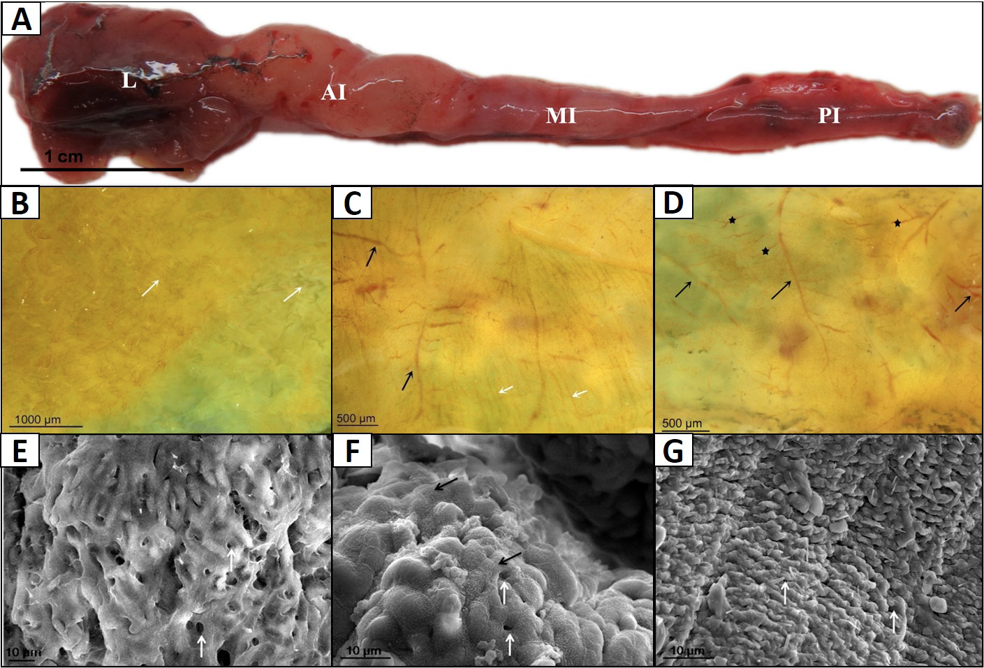
Fig 1
Morphology observation on the surface of the intestinal tract of P. dabryanus. A, intestinal tract of P. dabryanus; B, the anatomy of anterior intestine, the white arrow notes the intestinal folds; C, the anatomy of middle intestine, the white arrow notes the intestinal folds; the black arrow notes the blood vessels underneath the intestinal epithelial tissue; D, the anatomy of posterior intestine, black arrow indicate the main blood vessels underneath the intestine epithelial tissue, * notes the branching blood vessel network; E, the white arrow notes the secretory hole of the secretory cells; the black arrow indicate the microvillus; F, the white arrow notes the secretory hole of the secretory cells; the black arrow indicate the microvillus; G, the white arrow notes the secretory hole of the secretory cells; AI, anterior intestine; L, liver; MI, middle intestine; PI, posterior intestine