
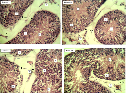
Figure 2
Photomicrograph of the section of the testes from T1, T2, T3 and T4. Note: seminiferous tubules are clearly separated and defined with thick layered spermatogenic cells (SC); testicular interstitium (TI) are clearly separated from the spermatogenic cells; spermatogenic cells are clearly distributed within the relatively spacious lumen (L); double arrow shows widen interstitial areas; arrow shows spermatogonial disruption (H & E x 400).