
Figure 2:
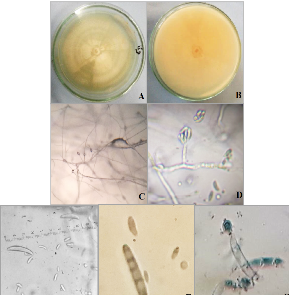
Neocosmospora rubicola (FCBP 1565). A: Colony front; B: Colony reverse: C: Conidial attachment with conidiophores under stereoscope; D: Conidial attachment under 4X magnification of microscope; E, F and G: Conidia.
Neocosmospora rubicola (FCBP 1565). A: Colony front; B: Colony reverse: C: Conidial attachment with conidiophores under stereoscope; D: Conidial attachment under 4X magnification of microscope; E, F and G: Conidia.