
Figure 3
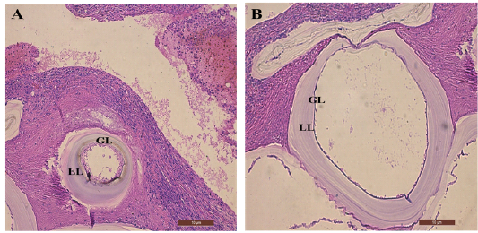
(A) and (B) show the secondary hydatid cyst in mice liver tissue of different sizes with (GL) germinal layer and (LL) laminated layer (10X), Stained with H&E. Scale bar = 10 μm
(A) and (B) show the secondary hydatid cyst in mice liver tissue of different sizes with (GL) germinal layer and (LL) laminated layer (10X), Stained with H&E. Scale bar = 10 μm