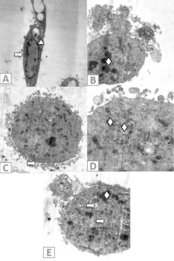
Fig 6
Ultrastructural changes of A549 cells. A, Untreated A549 cells: →, undamaged cell membrane; ∆, normal nuclear membrane. B, A549 cells exposed to ceranib-2 for 24 h. ◊, fragmented nucleus. C, →, Circular shaped cell and membrane blebbing. D, ◊, Lipid droplets in cells. E, ◊, DNA disintegration, →, holes in cell.