
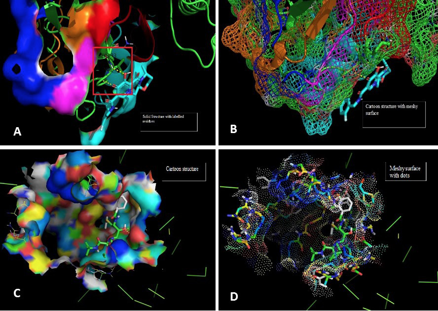
Fig. 6.
Molecular docking image analysis of Pol. The interactions are visualized between the protein and ligand of Dolutegravir with Pol protein. A shows the solid surface of the protein with residue labels Lysine and valine of amino acids. B represents the cartoon structure with meshy surface depicting the binding interaction of different residues of receptor with ligand atoms as shown. In figure C the cartoon structure and binding yellow lines show the hydrogen bonds. D demonstrates the meshy surface with dots.