
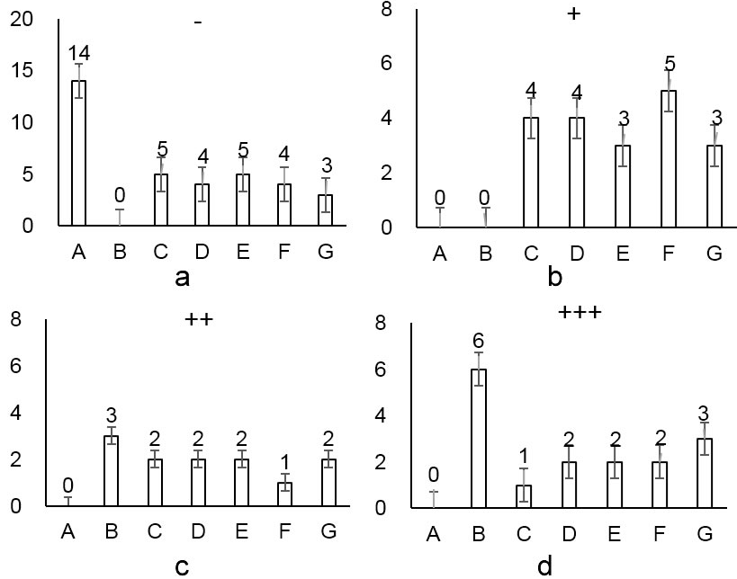
Effect of pathological changes in the cortex of brain tissue of mice with repeated cerebral ischemia-reperfusion. a: Cerebral cortical nerve cells are normal; b: Cerebral cortical individual nerve cell edema, individual neuron degeneration, cytoplasm light staining, fuzzy structure, individual neuron necrosis; c: Cerebral cortex a small number of nerve cell edema, scattered, a small number of neuronal degeneration The cytoplasm is lightly stained and the structure is fuzzy; d: Cerebral cortical nerve cells edema, most of the neurons are necrotic; A: Blank Group; B: Model Group; C: Nimodipine Group; D: Naoluotong Group; E: High-dose Spatholobus Grandiflprum Group; F: Mid-dose Spatholobus Grandiflprum Group; G: Low-dose Spatholobus Grandiflprum Group; -: Cerebral cortical nerve cells are normal; +: Cerebral cortical individual nerve cell edema, individual neuron degeneration, cytoplasm light staining, fuzzy structure, individual neuron necrosis; ++: Cerebral cortex a small number of nerve cell edema, scattered, a small number of neuronal degeneration The cytoplasm is lightly stained and the structure is fuzzy; +++: Cerebral cortical nerve cells edema, most of the neurons are necrotic.