
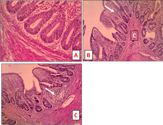
Figure 1
Intestine tissue sections of naturally IBDV infected chicken stained by H and E. A. Normal intestine tissue sections stained (x 200) B: Chicken intestine showing inflammatory cells infiltration of the mucosa (white arrow) and congestion (C) of the submucosa (x 100). C: Chicken intestine showing inflammatory cells infiltration of the mucosa (white arrow) (x 100).