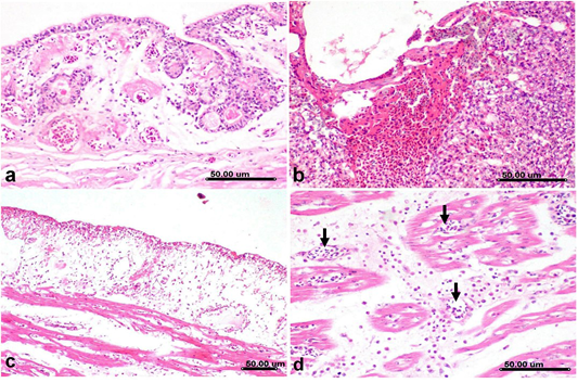
Histological sections of lung and heart from H5N8 infected duck, a) Primary bronchus showing deciliation of lining epithelium with congestion associated with interstitial edema and mononuclear cells infiltration. b) Parabronchus showing hemorrhage with proliferative reaction involving the air capillaries with mononuclear cells infiltration. c) Heart, epicardium showing desquamation of lining mesothelium associated with extensive subepicardial edema, congestion and mononuclear cells infiltration, note the extensive intermuscular edema of cardiac muscle fibers in the vicinity of subepicardial area. d) Myocardium showing necrosis of cardiac myocytes with mononuclear cells infiltration, congestion (arrow) and intermuscular edema. (Stain, H&E).