
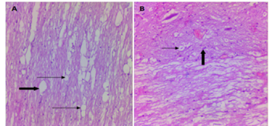
Figure 9
Micrograph of the longitudinal section of chitosan group at the site of the spinal cord injuries A. 8 weeks PO shows reduced cavities size and surround by glia cells (thin arrows) and moderate vacuolization (thick arrow). B. 16 weeks chitosan group shows small size cavity, injured area was filled with dense regenerative nerve fiber (thin arrow),