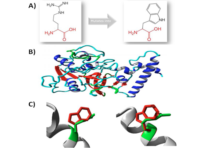
Fig 5
In silico examination of mutation and resultant protein: A, schematic structure of the original (left) and the mutant (right) amino acid; B, overview of the protein in ribbon-presentation. The protein is coloured by element; alpha helix, blue; beta strand, red; turn, green; 3/10 Helix, Yellow; random coil, cyan; C, Close up of mutation from two slightly different angles. The protein is coloured grey, the side chains of both the wild type and the mutant residue are shown and coloured green and red, respectively.