
Figure 4
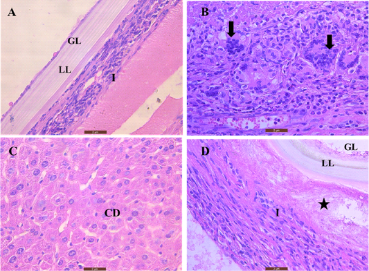
Sections of mice liver stained by hematoxylin and eosin (A and D) show the hydatid cyst layers: (GL) is the germinal layer, (LL) the laminated layer, and (I) infiltration of inflammatory cells that surround the hydatid cyst, the star indicates dead degenerated hepatic tissue (B) Shows accumulation of giant cells (arrows). (C) Illustrates cell degeneration (CD) and tissue necrosis adjacent to hydatid cyst (40X). Scale bar= 2µm