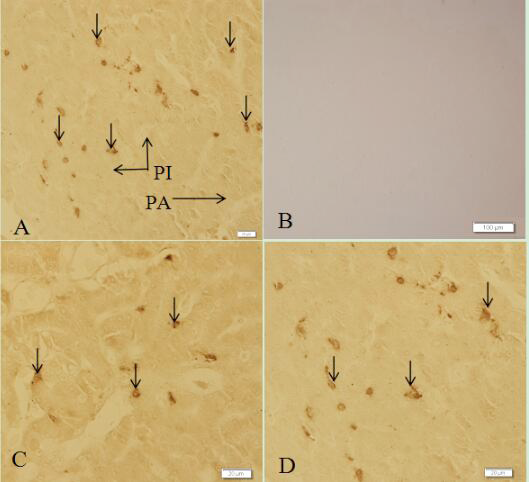
Fig 2
Histological structure of pancreas showing ghrelin-immunopositive cells in the pancreas of the African ostrich (SABC staining). A, a large number of ghrelin cells (arrows) were found within the pancreas; B, microphotograph of the absorption testing in the pancreas; C, ghrelin-ip cells (arrows) amound the Pancreatic acinar cells; D, ghrelin-ip cells (arrows) within the islets. PA, pancreatic acinus; PI, islet. (A, C and D) Scale bar: 20 μm; (B) scale bar: 100 μm.