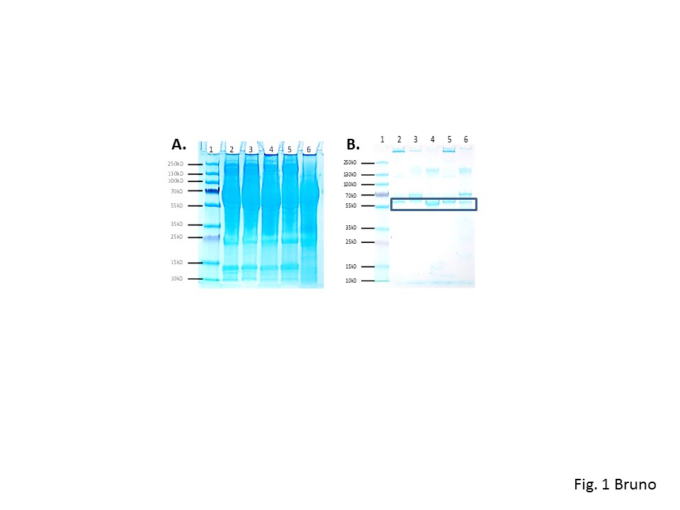
Figure 1
A. Coomassie blue-stained 4-20% polyacrylamide electrophoresis gel showing the typical appearance of murine antisera developed against R. rickettsii and R. typhi. B. Appearance of the same samples following removal of serum albumins, papain digestion and passage through a Protein A affinity column to remove Fc fragments. The remaing bands should represent Fab fragments of IgG (boxed at ~ 55 kD) or other isotypes (e.g., IgA or IgM) in the 4-20% PAGE gel.