
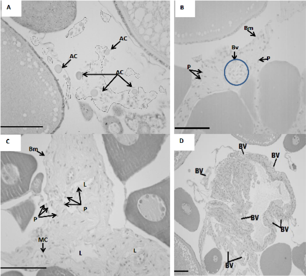
Fig. 5.
Apoptosis in POF. A, all irregular circulus show various type of blood cells. Arrows show apoptotic cells; B, arrow shows blood vessels. Piknosis were also detected; C, following inflection nuclei with condensed chromatin and showing pyknosis, were detected at the lumen of the follicle and at the follicular wall (arrows); D, blood vessels close to the follicular cells. Bm, basement membrane, MC, micropylar cell, AC, apoptotic cell; BV, blood vessels; P, pyknosis. Scale bars, 200µm. Stain: hematoxylin & eosin.