
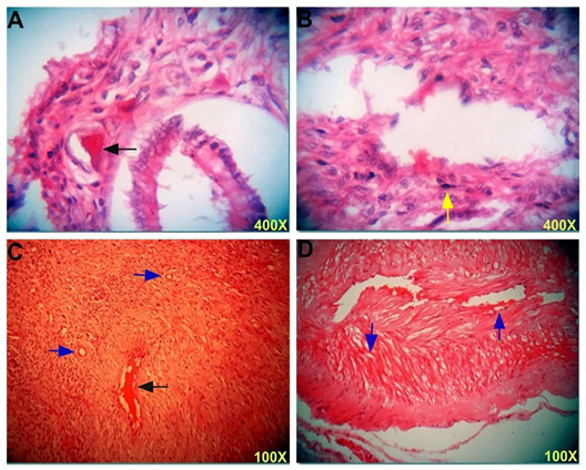
Figure 2
Section in the uterus post infection shows (A) severe congested blood vessels (black arrow) in the subserosal area. (B) Congested blood vessels with few mononuclear cells (yellow arrow) infiltration in the endometrium. (C and D) Congested of blood vessel (black arrow) and vacuolation (blue arrows) of muscle cells. H & E stain, 100X and 400X.