
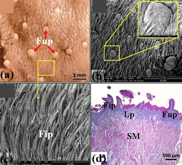
Fig. 1.
Light microscopical (a), scanning electron microscopic structure (b, c), and structure of the dorsal surface of the tongue of bear (d). Fup, fungiform papillae; red arrows, taste pores; Fip, filiform papillae; Lp, lamina propria; SM, striated muscles; yellow rectangles magnify the fungiform and filiform papillae in (b) and (c), respectively.