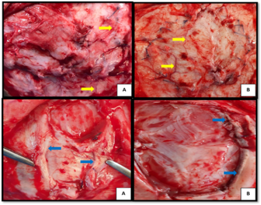
Figure 2
Macroscopic appearance of implantation sites, 2 weeks post-treatment of BTV treated hernias (A), and caprine ADM treated hernias (B), shows: numerous blood vessels (yellow arrows), deposition of white fibrous connective tissue in BTV more than caprine ADM (black arrows), incorporation of mesh with host tissue (blue arrows).