
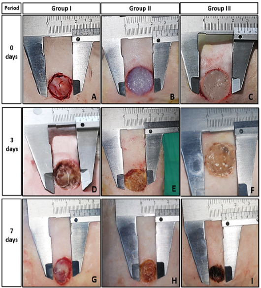
(A) The wound bed at zero day after debridement of group I. (B and C) The wounds of GII and GIII respectively at zero day, which are completely covered by fresh hydrogels. (D) The wound bed of GI at 3rd day shows the beginning of granulation tissue formation with swelling and rounding of wound edges. (E and F) The wounds defect of GII and GIII at 3rd day shows, no clear signs of inflammation, and the hydrogels become more homogenized with increased its adherence to the bed and edges of the wounds. (G) The wounds defect of control group at 7th still unfilled with granulation tissue, with a lesser reduction of wound area compared with GII and GIII. (H and I) The wounds of GII and GIII, respectively at 7th day post-treatment shows the organization and conversion of hydrogels to tissue-like structure.