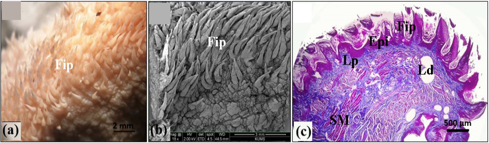
Fig. 2.
Light microscopical (a), scanning electron microscopic (b), and histological (Masson’s trichrome staining) of the lateral border of the tongue of bear (c). Lp, lamina propria; Ld, lipid droplets. For other abbreviations, see Fig. 1.