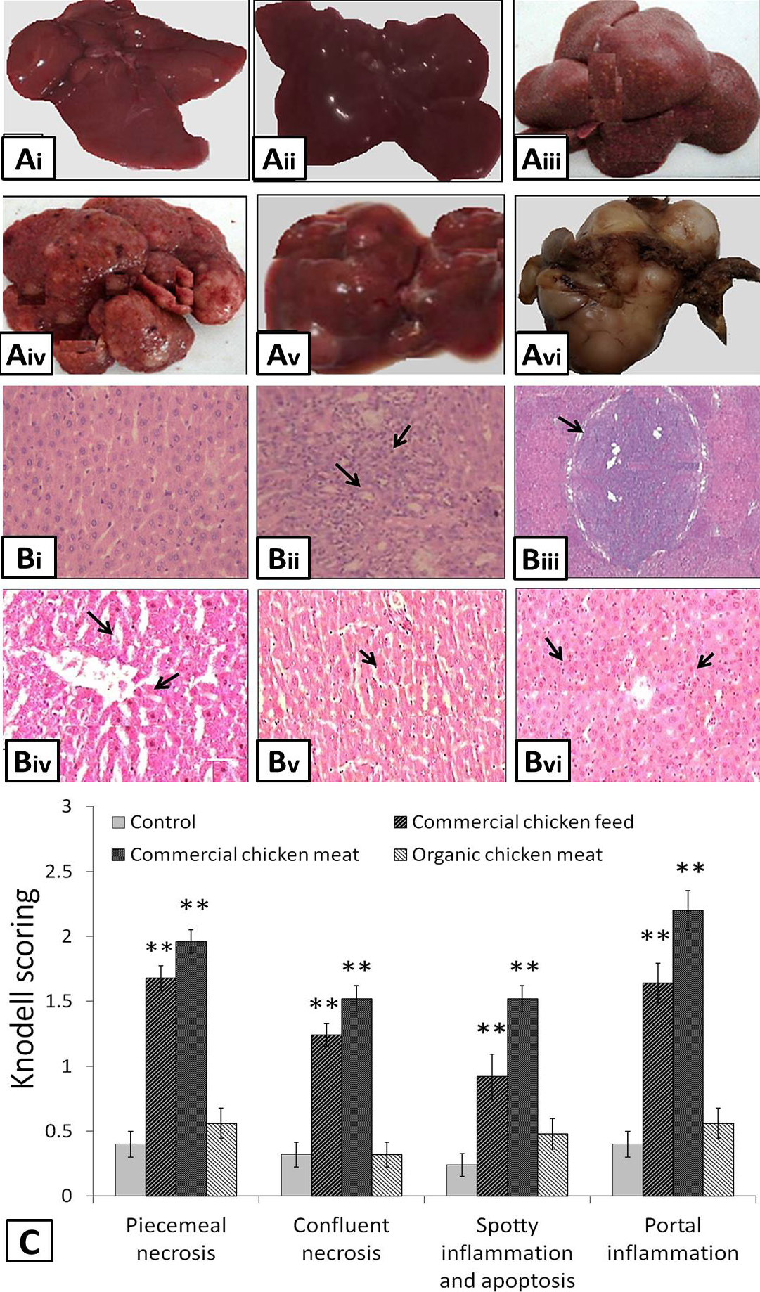
A, morphology of liver observed after the intake of commercial chicken feed, meat and organic meat. (i) and (ii) showed normal morphology of liver isolated from control and organic meat fed animals, (iii) inflamed, (iv) inflamed and nodular, (v) nodular and (vi) cirrhotic liver were seen in animals fed on commercial chicken feed and meat for six weeks. B, histopathological examination of liver samples showing the presence of (i) normal cells in control group A rats and organic chicken meat fed animals, (ii) Inflamed cells, (iii) cirrhotic nodule (iv) portal inflammation (v) confluent necrosis and (vi) spotty inflammation, necrosis with apoptosis were observed in liver cells isolated from the animals fed on commercial chicken feed and meat. C, Knodell scoring was done to assess the portal inflammation, necrosis, apoptosis and cirrhosis in liver samples. Values are mean ± SEM (n=25). **p<0.05 with respect to controls.
