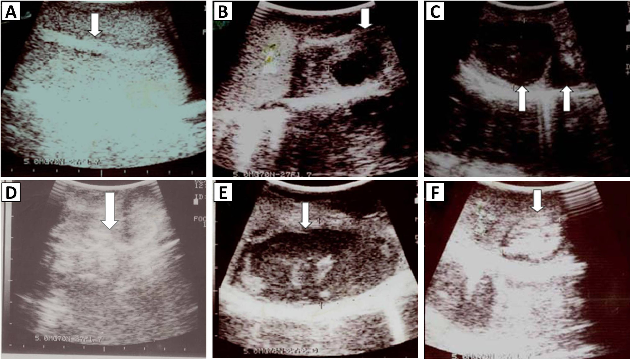
Longitudinal ultrasonograph of A, normal testis showing homogeneous and moderately echogenic parenchyma with hyperechoic mediastinum testis (arrow); B, showing sperm granuloma (arrow) at advance stage, having hyperechoic and anechoic areas; C, transverse ultrasonograph of the epididymal tail showing two sperm granulomata (arrows). Tissue below the lesion is hyperechoic, which is an ultrasound artifact known as enhanced through transmission; D, testis showing many hyperechoic areas scattered throughout the parenchyma (testicular mineralization); E, testicular abscess (arrow), showing hypoechoic and hyperechoic areas, with enhanced through transmission underneath the lesion (bright area); F, testis, showing a hyperechoic area surrounded by a hypoechoic line (arrow).