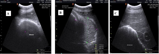
Figure 5
Ultrasonographic images of camel abomasum visualized from the right side from 8th-9th intercostal space using 3.5 MHz convex probe (longitudinal). (A) Before induction of acidosis, the abomasal wall appeared as distinct echogenic white line and the abomasal contents appeared hypoechoic and homogeneous. (B) After induction of acidosis, the wall appeared thicker than before induction of acidosis and the content appeared heterogenous in echogenicity and more echoic than before induction. (C) After treatment with Rumitone, the abomasal wall returned to distinct echogenic line and the content appeared hypoechoic and homogeneous.