
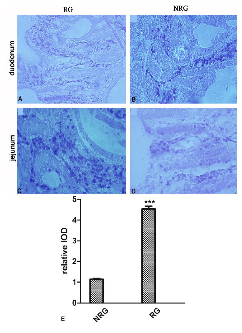
Fig. 1.
The distribution of IL2RB in the small intestine of sheep. Intestinal IL2RB expression in NRG and RG sheep was determined by immunohistochemical staining. A, C: Strong IL2RB positive expression was observed in T cells of mucosal layer and musclar layer. B, D: The positive signal of IL2RB was only detected in mucosal layer, especially in the lamina propria and epithelium. E: The average IOD was obtained by analysing IL2RB IHC in four random felds of each slide. Upper panel: representative image; lower panel: quantitative analysis. Results are presented as the mean± SEM (magnifcation, 200×, scale bars= 100μm).