
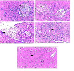
Figure 15
Photomicrograph of comparative figure of The normal group (group 1) showed a normal histological structure of the pancreas (Fig. 15a). The pancreas of diabetic control (group 2) showed severe necrosis and vacuolation of B-cells of Langerhans (Fig. 15 b), with extensive dilatation and congestion with inflammatory edoema (Fig. 15 c). The pancreas of group 3 showed haemorrhage with RBCs infiltration (Fig. 15 d). The pancreas of group 4 showed necrosis and vacuolation of pancreatic acini (Fig.15 e). (H&E., X 400).