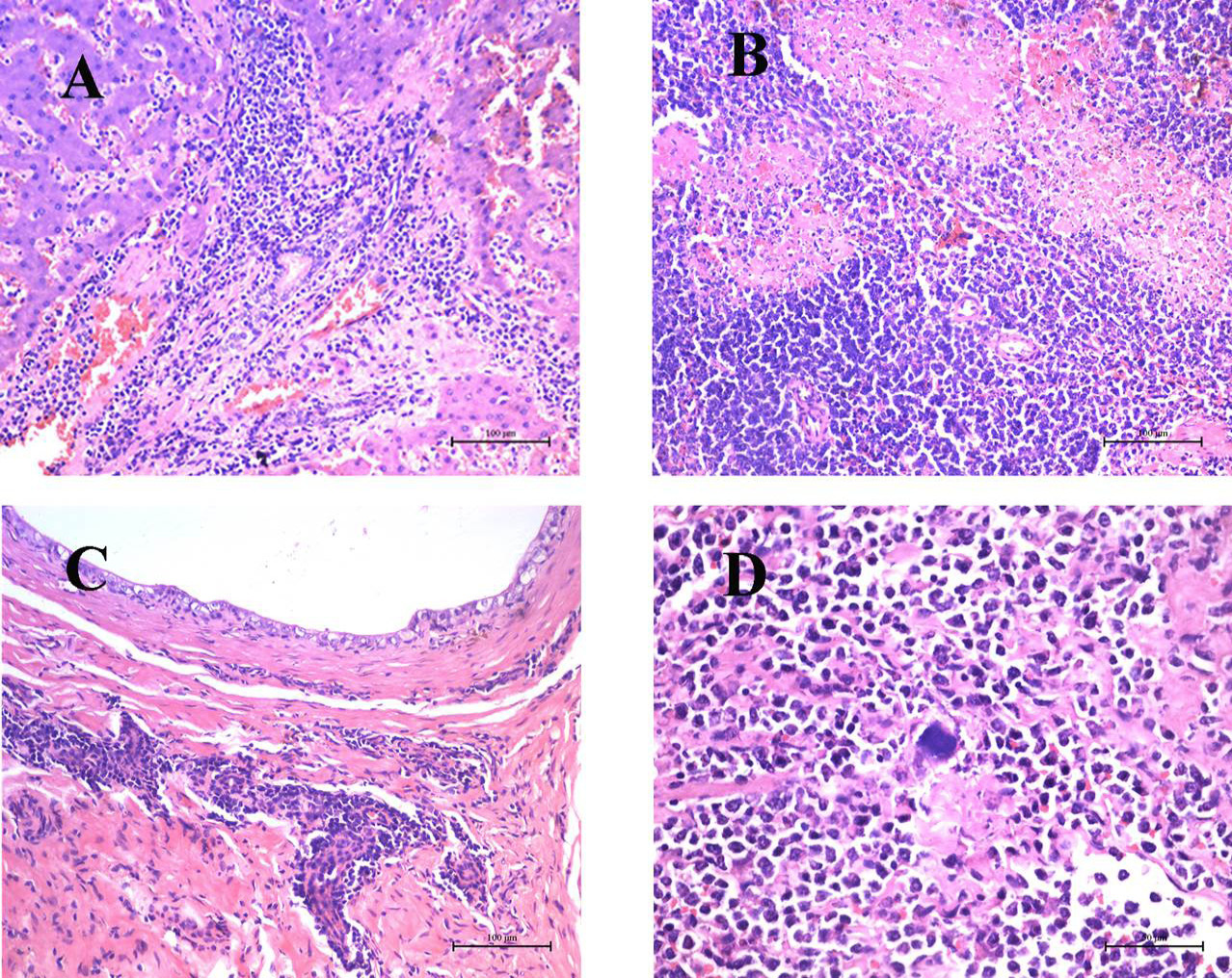
Pathologic lesions in liver, spleen, testicle and lymph nodes from the dog naturally infected with B. canis. A, liver showing severe infiltration of abundant lymphocytes and neutrophils in addition to hepatocellular necrosis and extensive vacuole degeneration of hepatocytes. HE Bar, 100 μm; B, spleen showing the presence of many granulomas with central necrotic areas in red pulp. HE Bar, 100 μm; C, testicle showing severe necrosis of spermatogenic cells and damaged seminiferous tubule structures accompanied by massive neutrophil, lymphocyte, and macrophage accumulation in interstitial tissue. HE Bar, 100 μm. D, lymph nodes showing proliferation of lymphocytes and reticuloendothelial cells along with deposition of fibrinous material; a bacterial bluish discoloration was found adjacent to the necrotic foci. HE Bar, 50 μm.