
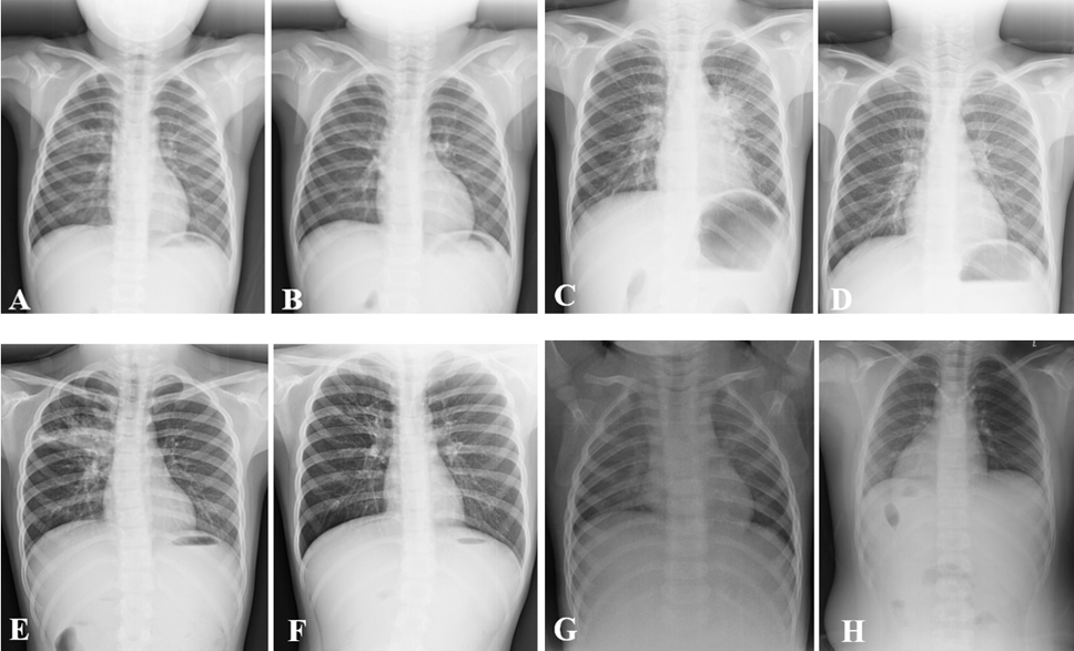
The Iconography features of Mycoplasma pneumoniae before and after treatment. A and B, Before and after the treatment of the patient (Light). C and D: Before and after the treatment of the patient (Severe). A, The obvious inflammatory infiltration accompanied with local patchy fuzzy shadow was found in the middle lobe of the right lung. B, The slight inflammation was residue after treatments. C, The patchy and fuzzy shadows could be seen in left hilar and left lower lung of the children with moderate mycoplasma pneumonia. D, Most fuzzy shadows in left hilar and left lower lung were markedly relieved. E, The interstitial inflammation was showed in double pulmonary and the clear atelectasis was also visual in the middle lobe of the right lung. F, Most of the interstitial inflammation was reduced, and the atelectasis in the middle lobe of the right lung was obviously disappeared. G and H were both as normal controls.