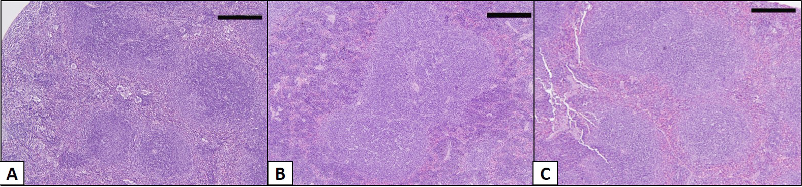
Figure 1
The effect of malarial infection on the distribution of red and white pulps in splenic tissue of female BWF1 mice. Hematoxylin and eosin (H/E) staining of spleen of lupus (A), live P. chabaudi (B) and gamma-irradiated P. chabaudi (C) infected group of BWF1 mice (n=10). The organs were removed at day 14 p.i. Sections of P. chabaudi infected groups have showed hypertrophied white pulps at the expense of red pulp. Scale bar = 100µm.