
Fig. 2.
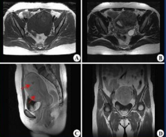
MRI examination (A, B, C and D show the examination results of uterine fibroids in different directions and the disease severity increases successively from A to D).
MRI examination (A, B, C and D show the examination results of uterine fibroids in different directions and the disease severity increases successively from A to D).