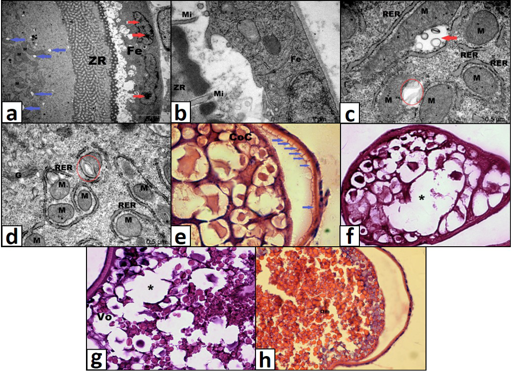
Lıght microscopic and electron micrographic structure of ovary of 2 mg/L TiO2 exposed group. A, chromatin condensation (red arrow) and cytoplasmic vacuolization (blue arrow); b, opening between zona radiata and follicular epithelium and degeneration at microvili structure; c, mitochondrial swelling (red arrow) and loss of organization of cristae (circle); d, loss of organization of cristae (circle); e, vacuolization under zona radiata (blue arrows); f, cytoplasmic vacuolization; g, unification of cortical alveoli (asterix); h, shrinking of mature oocyte. ZR, zona radiata; Fe, follicular epithelium; Mi, microvilli; M, mitochondria; G, golgi; RER, rough endoplasmic reticulum; CoC, cortical alveolus stage; Vo, vitellogenic oocyte; Om, mature oocyte. Magnification: e, f, g) x40, h) 20 x.