
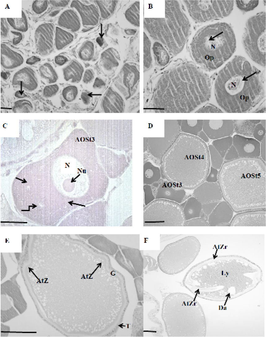
Fig. 1.
Pre-ovulatory atretic ovarian follicles. A, arrows show atretic oocytes as dark stain mass in the chromatin nucleolar stage; B, atretic oocytes at the perinocleolar stage, arrows show the development of clear spaces in the peripheral ooplasm; C, arrows show ealy vacuole formation; D, early phase of vascuolization, oocytes resorption; E, early degeneration of zona radiata in vitellogenic oocyte; F, fregmentation of zona radiata, follicular cells with protrusion extending into the oocyte cytoplasm. Ly, liquefaction of yolk granules; Da, degeneration of cortical alveoli; T, theca; G, granulosa; AtZr, atretic zona radiata. Scale bars: A, 30µm; B-F, 60 µm. Stain: hematoxylin & eosin.