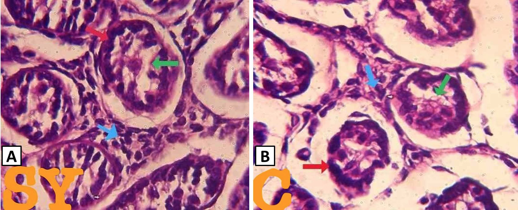
Fig. 2.
Histological structure of selenium yeast (SY) and control (C) demonstrating assembly of SFT including the BM (red arrow), germinal epithelium comprising of gonocytes and sertoli cells (green arrow) interstitial cells (blue arrow). Stain: H&E. Magnification: 100X.