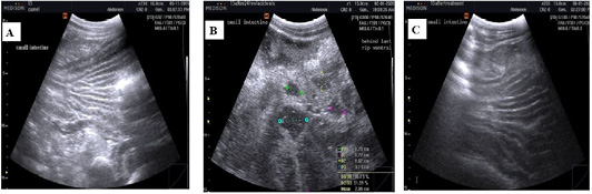
Figure 6
Ultrasonographic images of camel small intestine visualized low in the right paralumbar fossa using 3.5 MHz convex probe (longitudinal). (A) Before induction of acidosis: The intestinal contents were almost very hypoechoic and homogenous. (B) After induction of acidosis: Small intestine diameter is increased, the contents appeared heterogenous and more echoic compared to pre-acidosis induction. (C) After treatment with Rumitone: the intestinal contents returned to hypoechoic and homogenous.