Figure 5
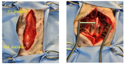
Photograph showing the initial steps of hemicordectomy. A. Shows the surgical incision over dorsal midline from L1 to L3. B. Elevated epaxial muscles from dorsal spinous processes, laminae, articular facets, and pedicles of L2 (arrow).