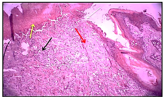
Figure 1
Histological section 3rdday of wound healing control group showed the non – epithelialization surfaces (white arrow), collagen fibers proliferation forming a matrix (red arrow). Granulation tissue formation (black arrow) with the presence of red blood cells (yellow arrow). (Hematoxylin and Eosin ,10 X).