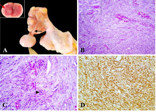
Figure 1
Adult Granulosa cell tumor, ovary, camel. (A) Firm grayish white nodule attached to the ovary by a long stalk (black arrow), Inset, hemorrhagic cut section. (B) Neoplastic cells arranged in solid sheets, supported by fine fibrovascular stroma, HE x 200. Inset, round to ovoid uniform nuclei with pale chromatin and coffee-bean-like nuclear groove, HE x 400. (C) Sarcomatous change characterized by pleomorphic cells with irregular, hyperchromatic, bizarre nuclei (arrowhead) showing variable mitosis, HE x 400. (D) Neoplastic cells expressed cytoplasmic positive reaction for vimentin, x 200.