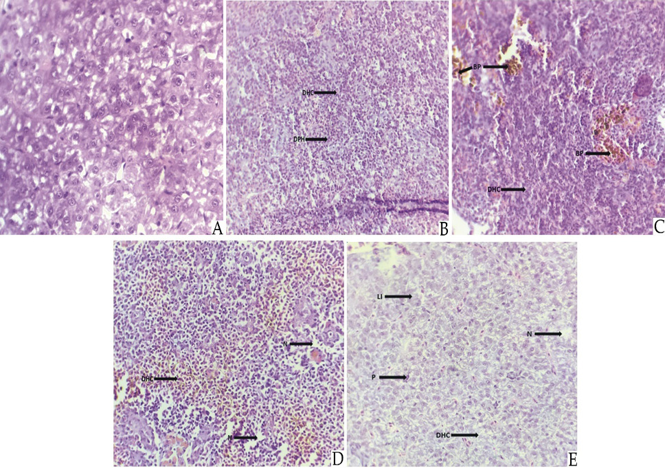
Fig. 8.
Effect of different concentration of cypermethrin on various histological structure of Liver of Schizothorax esocinus. (A) Control showing normal hepatocytes, hepatopancrease and sinusoids. (B) fish exposed to 1 ppb showing (DHC) Disappearance of hepatic cell wall and (DPH) Densly packed hepatocytes. (C) fish exposed to 2 ppb showing (BP) Bile pigment and (DHC) Disappearance of hepatic cell wall. (D) fish exposed to 3 ppb showing (DHC) Disappearance of hepatic cell wall and (N) Necrosis. (E) fish exposed to 4 ppb showing (Li) Leukocyte infiltration, (DHC) Disappearance of hepatic cell wall, (P) Pyknosis and (N) Necrosis.