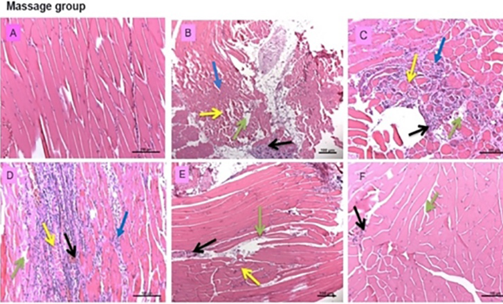
Fig. 4.
Microstructure of tibialis anterior in massage only group (H&E staining) A, normal tissue (pre-injury); B-F, the injured tissues at day 2(B), day 5(C), day 8 (D), day 12 (E) and day 16 (F). Yellow arrows, myofiber arrays; Black arrows, myocytes; Geen arrows, connective tissues; Blue arrows, satellite cells.