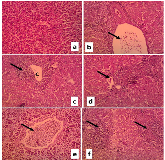
Liver sections of duckling naturally infected with DVH (H&E, X100) showing
a. Normal liver tissue
b. Portal vein and bile duct congestion (arrow) accompanied by hepatic cord disorganization
c. Focal area of coagulative necrosis (C) characterized by inflammatory cells infiltration (arrow) accompanied byhepatic cord disorganization and vacuolar degeneration in the cytoplasm of the hepatocytes
d. Focal area of coagulative necrosis characterized by inflammatory cells infiltration (arrow) accompanied byhepatic cord disorganization and vacuolar degeneration in the cytoplasm of the hepatocytes
e. Diffuse vacuolar degeneration and severe necrosis of hepatocytes and lymphocytic infiltration of the central vein
f. Multifocal area of coagulative necrosis characterized by inflammatory cells infiltration (arrow) accompanied byhepatic cord disorganization and vacuolar degeneration in the cytoplasm of the hepatocytes