
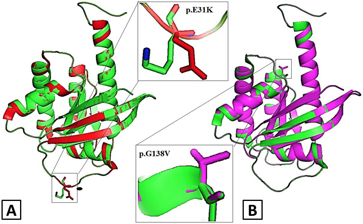
Fig. 3.
Structural alignment of wild-type and mutant models of KRAS in cartoon mode visualized by PyMol (A) p.E31K KRAS mutant model (red) superimposed with native KRAS protein (green) (B) p.G138V KRAS mutant model (magenta) superimposed with native KRAS protein (green) KRAS. Rectangle boxes depict the zoomed-in view of mutation sites.