
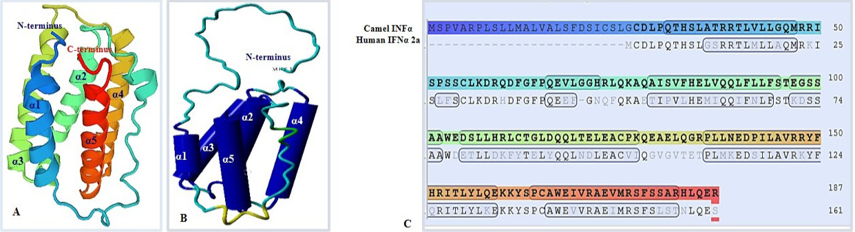
Fig. 6.
Predicted 3D structure of C. dromedarius IFNα protein shows the overall secondary structure in ribbon form (A) and cartoon form (B). Alpha helices are labeled from α1 to α5. (C) Model-template alignment of amino acid residues of C. dromedarius IFNα and Homo sapiens IFNα2a. Components of the secondary structure are shown in blue (α helices) and brown (coils). Identical amino acid residues are shown in bold black.