
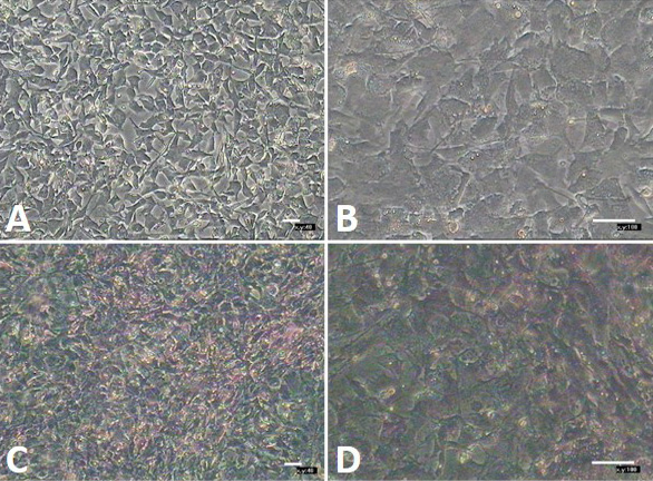
Fig. 11.
Hepatocyte induced differentiation of CAFSCs in vitro. A and B, morphology observation of induced cells under inverted microscope, cells present polygonal and foliated after culture for 12 d; C and D, Glycogenosome present amaranth by glycogen staining. This can prove that high-passaged CAFSCs (P25) still maintain induced differentiation potential.