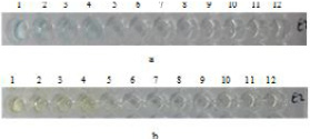
Fig. 10.
HCV3a E2 antibodies confirmation after ELISA. Wells 1-12 contain different dilutions ranging from 100% pure to 10 times serially diluted E2 antibodies. (a) Blue color after adding TMB substrate. (b) Color change from blue to yellow after adding 12.5% H2SO4 as stop solution.