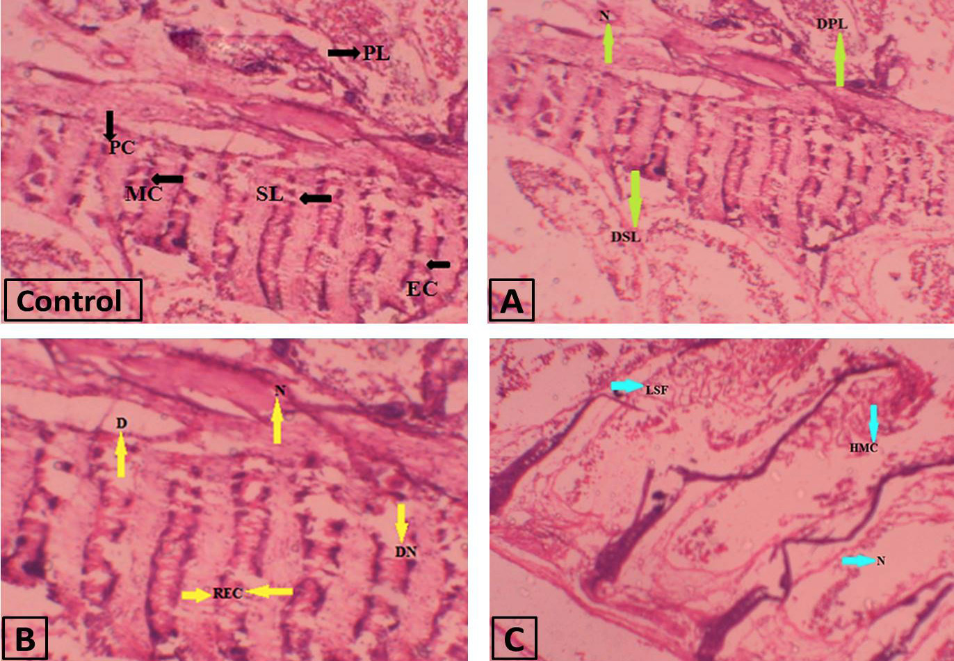
Figure 2:
Photomicrograph of Gills of Hypophthalmicthyes molitrix. section of control group showing primery lamella (PL), secondary lamellae (SL), mucous cells (MC), epithelial cells (EC) and pillar cells (PC). Group A showing necrosis (N), damaged secondary lamella (DSL), damaged primary lamellae (DPL). Group B showing necrosis (N), degeneration (D), damaged nuclei (DN) and rupturing of epithelial cells (REC). Group C showing necrosis (N), hypertrophy of mucous cells (HMC) and lamellar severe fusion (LSF).