
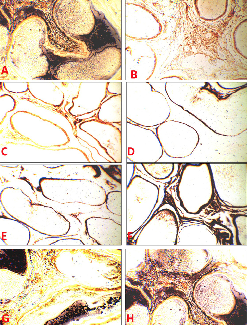
Fig. 5.
(A) Tubuli recti of the control group showing normal reticular fibers. (B, C, D and E) Tubuli recti of M, AL, AM and AH group showing degeneration and fragmentation of reticular fibers (F, G and H) Tubuli recti of ALM, AMM and AHM group showing the normal appearance of reticular fibers. (Silver stains x200).