
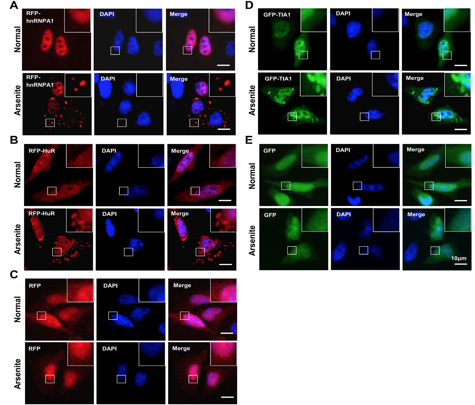
Fig. 1.
Image data for SG formation and nucleus/cytoplasm localization of hnRNP A1, HuR and TIA1 upon oxidative stress. HeLa cells were cultured and transiently transfected with plasmids of RFP-hnRNP A1 (A), RFP-HuR (B), RFP vector(C), GFP-TIA1 (D), and GFP vector (E), respectively, as indicated. After 24 hours, cells were untreated (Normal) or treated with 0.5 mM sodium arsenite for 1 hour (Arsenite). The nuclenus section was stained by DAPI. An inverted research microscope Leica was then used to collect the image data. Bar=10 μm.