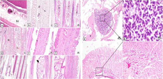
Photomicrograph of gills: Marked diffuse edema and severely congested blood vessels are seen in gill arch (a). Multifocal lamellar telangiectasia is noticed in affected gills (b). Heavy eosinophilic granular cells infiltration with variable number of inflammatory cells (c). Hyperplasia of the secondary gill lamellae admixed with necrosis of adjacent gill elements (d). Sever destruction and necrosis in the secondary gill lamellae with inflammatory cells infiltration (e-g). Attached isopod spp. (arrow) to the surface of gills with intense inflammatory reaction (h-i). Edema (Ed), congestion (Cong), telangiectasia (T), necrosis (N), hyperplasia (Hy), eosinophilic granular cells (E). Photomicrograph of subcutaneous and intestine of fish: Subcutaneous tissue showing Myxosporidian cysts with higher magnification showing numerous myxobolus spores (j-k). Intestine of fish showing edema of mucosa associated with intense inflammatory cells infiltration in the sub epithelial layer (l). Intestine of fish showing higher power of inflammatory cells infiltration with numerous numbers of eosinophilic granular cells (m).