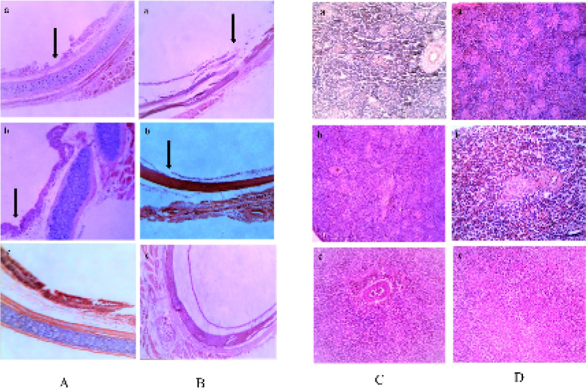
Fig. 3.
(A and B) Histopathological lesions observed in trachea of Japanese quail (A) and myna (B) were mainly discontinuation of surface epithelium. Loss of cilia of the lining epithelium and disturbance of coherence among various histologic layers were also noticeable. (C and D) Histopathological lesions observed in spleen of Japanese quail (C) and myna (D) were mainly enormous infiltration with mononuclear lymphocytes, especially in the area of red pulp. This region also showed hyperemic changes, petechial hemorrhages were also observed.