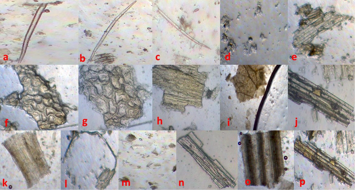
Figure 6
Powder drug microscopy of Oxalis pes-caprae (Microphotography).
Key: a, b, c, unicellular and multicellular trichomes; d, calcium oxalate crystals; e, columnar palisade cells; f, epidermis with palisade cells; g, fragment of epidermal cells; h, fragment of ground tissue; I, fragment of leave showing vein islet; j, needle shape crystals; k, parenchyma cells; l, phloem fibers; m, starch grains; n, vessels with annular thickening; o, xylary trachieds; p, xylem vessels.