
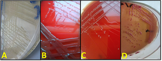
Figure 3
A: Growth of P. multocida on BHI agar plate, B: growth of M. haemolytica on blood agar plate with β-zone of hemolysis, C: growth of P. multocida on blood agar plate with no hemolysis, D: growth of P. multocida on MacConkey agar plate with lactose fermentation.